Leg Tendon Chart / Ankle Anatomy Muscles And Ligaments. An upper arm muscle composed of 2 parts, a long head and a short head. 16″ 18″ just below knee or hock to pastern (k) 10″ 12″ 14″ 16″ 18″ mesh sheet and turnout blanket sizing chart. Reflexes help to maintain proper muscle tone, balance, and responsiveness of the legs and feet to stimuli such as stepping on a sharp object. Repeat and compare to the other leg. A muscle of the medial thigh that originates on the pubis.
Soleus upper fibula, soleal line of tibia: Flexes hip and draws leg forward. As much as you may have wanted to see them here, leg extensions and curls didn't make the cut for this list of the 10 best leg exercises. It pulls the leg toward the body's midline (i.e. Calcaneum (by achilles tendon) raises heal when leg is straight.
Diagram Showing The Tendons And Ligaments Of The Ankle And Foot Download Scientific Diagram from www.researchgate.net In the leg muscles diagram above, there are many muscles that make up your legs and support it to move. For women, shaping the thigh muscles is an essential goal of physical fitness. The muscles of the leg anatomy chart shows in every possible view the way that the muscles and other pieces of the leg work together in motion and flexibility. Using the muscle and nerve chart let's say you're interested in knowing all the muscles innervated by the ulnar nerve. The nerve signals in these reflexes come from stretch receptors located in the joints, ligaments, tendons, and even the muscles themselves. It acts as a tensor of the arches of the foot, but can also be added with the first digit and plantar flexion of its first phalanx. An upper arm muscle composed of 2 parts, a long head and a short head. Pain above the knee cap (yellow).
16″ 18″ just below knee or hock to pastern (k) 10″ 12″ 14″ 16″ 18″ mesh sheet and turnout blanket sizing chart.
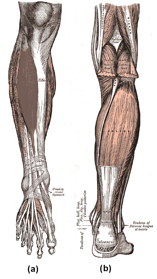
All of these tendons protect and house the four ligaments inside of your knee, including your medial collateral ligament, lateral collateral ligament, anterior cruciate ligament and posterior cruciate ligament. The knee jerk reflex is mediated by the l3 and l4 nerve roots, mainly l4. The achilles tendon attaches the muscles of the calves to the bones of the ankle and foot. However, many reflex pathways are also active in the legs and foot. symptoms tendonitis causes pain that increases with activity or stretching of the. The muscles that make up the quadriceps are the strongest and leanest of all muscles in the body. The gastrocnemius is the larger calf muscle, forming the bulge visible beneath the skin. It inserts onto the linea aspera of the femur. With the lower leg hanging freely off the edge of the bench, the knee jerk is tested by striking the quadriceps tendon directly with the reflex hammer. Tendons that make this possible include: Repeat and compare to the other leg. The hamstring tendon connects the hamstring to the knee. Tibial nerves stimulate muscles in the back of the lower leg.
If the tendon becomes inflamed because of overuse a sharp pain is felt at the back of the knee. In the leg muscles diagram above, there are many muscles that make up your legs and support it to move. The first diagram summarizes the different muscular compartments (fascial compartments) of the thigh and leg, and the different fascias (crural fascia, intermuscular septum, interosseous membrane, adductor canal, fascia lata) The myology of the lower limb is also particularly well represented in this atlas of anatomy, with multiple anatomical charts and diagrams: Leg muscle chart muscles of the leg origin insertion action rectus femoris anterior inferior iliac spine base of patella knee extension/hip flexion anterior superior iliac spine superior aspect of medial surface of tibial shaft knee flexion & laterally rotates hip joint gluteus maximus upper portion of ilium (sacrum & coccyx) gluteal tiberosity.
Muscle Charts Massagelongbeachca Com from www.massagelongbeachca.com Movement at the hip joint occurs when you bend backwards and forwards, and when you swing your leg while walking. It is controlled by the obturator nerve. Leg muscle chart muscles of the leg origin insertion action rectus femoris anterior inferior iliac spine base of patella knee extension/hip flexion anterior superior iliac spine superior aspect of medial surface of tibial shaft knee flexion & laterally rotates hip joint gluteus maximus upper portion of ilium (sacrum & coccyx) gluteal tiberosity. A pulled muscle in your leg will likely result in localized pain in the affected muscle. But let's be honest, you want to maintain the muscle definition in your legs all year round—whether you're wearing a pair of swim trunks or the perfect pair of denim jeans. Nothing says i hit the gym hard like a set of ridiculously sculpted quads and calves—especially during beach season. You may also find that the muscle is swollen and tender to the touch. Plantarflexion of foot at ankle, flexion of knee:
The hamstring tendon connects the hamstring to the knee.
The achilles tendon attaches the muscles of the calves to the bones of the ankle and foot. Calcaneum (by achilles tendon) raises heal when leg is straight. The hamstring tendon connects the hamstring to the knee. The nerve signals in these reflexes come from stretch receptors located in the joints, ligaments, tendons, and even the muscles themselves. Height (j) 5.5″ 6.5″ therapeutic no bow leg wraps 12″. In order to achieve the best results from your ireliev device, you should follow proper tens and ems muscle stimulation pad placement pictographics. Soleus upper fibula, soleal line of tibia: Included are more than a dozen illustrations like the vastus lateralis, adductor brevis, rectus femoris, semi tendinosus and many, many more. A muscle of the medial thigh that originates on the pubis. The hamstring is one of three muscles at the back of the leg between the hip and the knee. It acts as a tensor of the arches of the foot, but can also be added with the first digit and plantar flexion of its first phalanx. Tendons that make this possible include: Then next one, further down, looks at pain behind the knee.
#muscle and tendon pain in legs #muscles and tendons of the leg and foot #muscles and tendons of the lower leg #muscles ligaments and tendons of the lower leg #muscles tendons and ligaments of the upper leg It is controlled by the obturator nerve. Master these leg exercises to pack muscle and size onto your glutes, quads, and hamstrings. The gastrocnemius is the larger calf muscle, forming the bulge visible beneath the skin. Leg muscle chart muscles of the leg origin insertion action rectus femoris anterior inferior iliac spine base of patella knee extension/hip flexion anterior superior iliac spine superior aspect of medial surface of tibial shaft knee flexion & laterally rotates hip joint gluteus maximus upper portion of ilium (sacrum & coccyx) gluteal tiberosity.
9 9d Muscles That Cause Movement At The Ankle Medicine Libretexts from s3-us-west-2.amazonaws.com By lee boyce and ebenezer samuel, c.s.c.s. Included are more than a dozen illustrations like the vastus lateralis, adductor brevis, rectus femoris, semi tendinosus and many, many more. An upper arm muscle composed of 2 parts, a long head and a short head. But let's be honest, you want to maintain the muscle definition in your legs all year round—whether you're wearing a pair of swim trunks or the perfect pair of denim jeans. Then next one, further down, looks at pain behind the knee. The myology of the lower limb is also particularly well represented in this atlas of anatomy, with multiple anatomical charts and diagrams: Pain above the knee cap (yellow). The calf muscle, on the back of the lower leg, is actually made up of two muscles:
The nerve signals in these reflexes come from stretch receptors located in the joints, ligaments, tendons, and even the muscles themselves.
Height (j) 5.5″ 6.5″ therapeutic no bow leg wraps 12″. The term pulled muscle typically refers to a muscle strain and happens from overexertion and poor flexibility. The hamstring tendon connects the hamstring to the knee. If the tendon becomes inflamed because of overuse a sharp pain is felt at the back of the knee. Leg muscle chart muscles of the leg origin insertion action rectus femoris anterior inferior iliac spine base of patella knee extension/hip flexion anterior superior iliac spine superior aspect of medial surface of tibial shaft knee flexion & laterally rotates hip joint gluteus maximus upper portion of ilium (sacrum & coccyx) gluteal tiberosity. Hind (d) 10″ 10.5″ royal fetlock boots: The largest muscle masses in the leg are present in the thigh and the calf. Included are more than a dozen illustrations like the vastus lateralis, adductor brevis, rectus femoris, semi tendinosus and many, many more. Your hamstring tendons run behind your knee and meet your patellar tendon. It is controlled by the obturator nerve. Soleus upper fibula, soleal line of tibia: Tibial nerves stimulate muscles in the back of the lower leg. Based on the body part you're looking to train, these links will take you to page that will specify an optimal way to position yourself and the electrode pad during stimulation.
Leg, ankle, foot, posterior thigh gastrocnemius medial epicondyle of femur, lateral epicondyle of femur: leg tendon. A muscle of the medial thigh that originates on the pubis.
Share :
Post a Comment
for "Leg Tendon Chart / Ankle Anatomy Muscles And Ligaments"


Post a Comment for "Leg Tendon Chart / Ankle Anatomy Muscles And Ligaments"